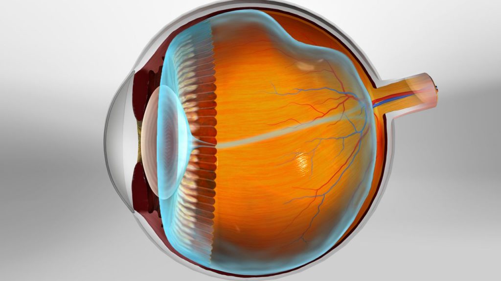
Posterior Vitreous Detachment (PVD)

Posterior Vitreous Detachment (PVD)
Posterior Vitreous Detachment (PVD) is a common and naturally occurring phenomenon in the eye, and while it typically does not pose a significant threat to eye health, it can result in visual disturbances. It is crucial to delve deeper into the understanding of PVD, exploring its multifaceted causes, its diverse array of symptoms, and the various management options available. By gaining a comprehensive grasp of this eye condition, individuals can take proactive steps to ensure their vision remains clear and healthy, thus enhancing their overall quality of life.
What is Posterior Vitreous Detachment (PVD)?
Causes
The primary cause of PVD is the aging of the vitreous, which becomes less gel-like and more liquid with time. This change leads to the vitreous gradually pulling away from the retina. Factors that may increase the risk of PVD include:
- Age (more common in individuals over 50)
- Nearsightedness (myopia)
- Eye injuries or traumas
- Previous eye surgeries
- Symptoms of PVD
Risk Factors
As the vitreous separates from the retina, individuals may experience various visual symptoms, such as:
- Floaters or specks in the field of vision
- Flashes of light, especially in peripheral vision
- The sensation of a curtain or cobweb-like veil across your field of vision
- While PVD is often a harmless and age-related process, sudden flashes of light or a significant increase in floaters should be examined by an eye care specialist to rule out any associated retinal issues.
Symptoms of Posterior Vitreous Detachment (PVD)

Symptoms of Posterior Vitreous Detachment (PVD)
You might also notice the presence of intermittent flashing lights, resembling faint flickers at the outer periphery of your vision. While these lights typically do not signal a significant issue, it is imperative to undergo a comprehensive eye examination, as there is a rare possibility of a retinal tear or detachment, which demands timely attention.
Treatment and Assessment
Posterior vitreous detachment typically does not necessitate any specific treatment. In most cases, floaters tend to naturally resolve within a few months, although occasional residual floaters may persist. Over time, you’ll likely become less aware of them and adapt to their presence.
During the examination, your pupils will be dilated using eye drops. The medical professionals will carefully inspect the periphery of the retina to detect any signs of a retinal tear. Various instruments may be employed to perform these examinations, and in some instances, a special contact lens may be utilized, along with gentle external pressure applied to the outer part of the eye for a more thorough evaluation.
To learn more about managing PVD or to schedule an eye examination, please reach out to us:
